Images
All images are copyrighted by Mayo Clinic unless otherwise noted.
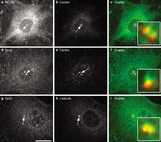
 Dyn2 localizes to the pericentriolar material and centrioles
FR cells were co-stained for endogenous Dyn2 using either the anti-pan-dynamin antibody MC-63 (a) or the anti-Dyn2 antibody (d, g) and the centriolar protein centrin (b, e) or the centrosomal protein γ-tubulin (h). Overlays also are shown (c, f, i). Insets in c, f and i show enlargements of the boxed centrosomal regions. Note that Dyn2 (green) is beside the centrin-positive spots (red, f), whereas in there is extensive co-localization (i)between Dyn2 (green) and γ-tubulin (red). Arrows indicate the centrosomal localization of Dyn2, centrin and γ-tubulin.
Source: Thompson, Heather M., et al. Dynamin 2 binds gamma-tubulin and participates in centrosome cohesion. Cell Biology, 2004; doi.org/10.1038/ncb1112. Used under Creative Commons license. https://creativecommons.org/licenses/by-nc-sa/4.0
 Dynamin co-localizes with cortactin in the lamellipodia of growth factor-stimulated fibroblasts
Dyn2 (green) and cortactin (red) merge into large cortical ruffles (arrows) situated at the leading edges of the actively migrating cells.
Source: Mark A. McNiven, et al. Regulated interactions between dynamin and the actin-binding protein cortactin modulate cell shape. Journal of Cell Biology, 2000; doi.org/10.1083/jcb.151.1.187. Used under Creative Commons license. https://creativecommons.org/licenses/by-nc-sa/4.0
 HBV infected cells secrete CD63 positive exosomes
HepG2.2.15 cells are stably infected with HBV and secrete CD63 positive exosomes (green), which can also contain hepatitis B virions, as they migrate.
 The antiviral protein MxB induces changes to mitochondria
HeLa cells were transfected for 48 hours with (a) GFP-vector alone or (b) MxB K131A-GFP prior to fixation and viewing by electron microscopy Normal cristae are observed in the mitochondria of the vector-only control cells compared with that of the cells expressing the mutant protein, which possess largely hollowed organelles with very few residual cristae. Hollow mitochondria were not observed in any treated control cells.
Nature Communications. 2020; doi:10.1038/s41467-020-14727-w. Used under Creative Commons license. https://creativecommons.org/licenses/by-nc-sa/4.0
 Hepatocytes possess size-based LD subpopulations
Primary rat hepatocytes possess a second population of much smaller lipid droplets (LDs) encased within degradative lipophagic vesicles resembling autolysosomes-late endosomes. Numerous dark granules indicative of lysosomal content are observed in close contact to the LDs.
Source: Schott, Micah B., et al. Lipid droplet size directs lipolysis and lipophagy catabolism in hepatocytes. Journal of Cell Biology, 2019; doi.org/10.1083/jcb.201803153. Used under Creative Commons license. https://creativecommons.org/licenses/by-nc-sa/4.0
 TEM image of activated MVBs containing viral antigens in HBV-expressing cells
Lower magnification transmission electron microscopy (TEM) field from a HepG2.2.15 cell in which multivesicular bodies (MVBs) and tubules are highlighted in red, demonstrating the high frequency of MVB tubulation in the virus-producing cells.
Source: Inoue, Jun, et al. HBV secretion is regulated through the activation of endocytic and autophagic compartments mediated by Rab7 stimulation, Journal of Cell Science, 2015; doi.org/10.1242/jcs.158097. Used under Creative Commons license. https://creativecommons.org/licenses/by-nc-sa/4.0