Vesicular Trafficking, Cytoskeletal Dynamics and Signaling in Acinar Cells
Research in the Pancreatitis and Acute Outcomes Laboratory has determined the role of the nonreceptor tyrosine kinase c-Src in the maturation and packaging of the enzyme amylase into zymogen granules in pancreatic acinar cells. This work was published in Scientific Reports, 2018.
Our current studies explore the mechanisms underlying the normally apically polarized secretion from the pancreas under physiological conditions, and also the pathological basolateral release that takes place during pancreatitis. This basolateral release is responsible for the increase in circulating levels of amylase and lipase during pancreatitis, which is used to diagnose the disease. This basolateral release into the visceral adipose may also be a crucial step in identifying worsening pancreatitis outcomes.
Pathophysiology of systemic injury in acute pancreatitis
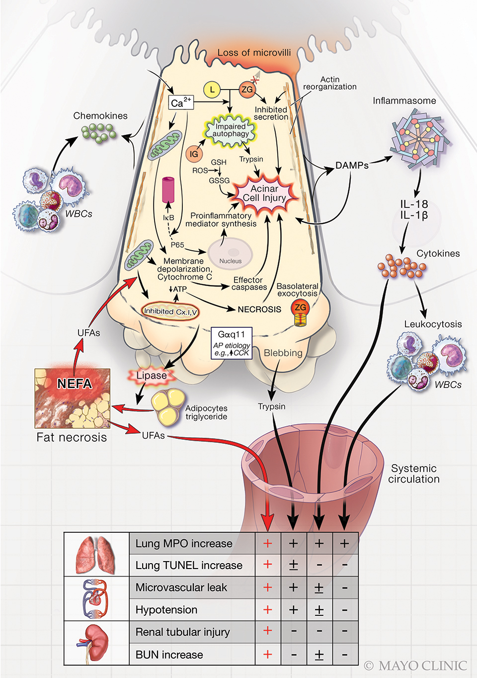
Role of the exocrine pancreas in systemic injury during acute pancreatitis.
Key
- Adipo. TG — Adipocyte triglyceride
- BUN — Blood urea nitrogen
- DAMP — Damage-associated molecular patterns
- L — Lysosome
- MPO — Myeloperoxidase
- NEFA — Nonesterified fatty acids
- UFAs — Unsaturated fatty acids
- ZG — Zymogen granule
During injury to an exocrine acinar cell, products released play a role in multisystem injury. The injurious signaling shown is modeled by high-dose cholecystokinin (↑CKK), commonly used to cause mild, self-limited acute pancreatitis.
The + indicates the components of systemic injury directly induced by the agent projecting the arrows based on at least two independent publications. For example, unsaturated fatty acids (UFAs) can cause all components of multisystem organ failure. The ± indicates where the evidence is indirect or unclear. The ‒ indicates lack of evidence.
This image shows a normal pancreatic acinus under basal state on the left. The image on the right shows how zymogen granules (red) are released basolaterally (arrows) from the acinus after exposure to a stimulus causing pancreatitis.
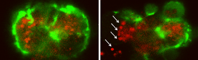 Pancreatic acinus before and after exposure to pancreatitis causing stimulus
Images from Dr. Singh's lab show how zymogen granules (red) are released basolaterally (arrows) from the acinus after exposure to a stimulus causing pancreatitis.
This approach, complemented by genetic and pharmacologic manipulation of the signaling molecules, along with biosensors that show their activation status, allows us to visualize the cell biology of acini during health and disease, in real time. These methods can also be applied to other relevant cells (such as inflammatory cells) involved in pancreatitis.