Videos
Videos and gifs from Dr. Lerman's Renovascular Disease Lab demonstrate imaging of renal structure and function, providing good representation of the disease models we study and our focus on MRI and CT imaging.

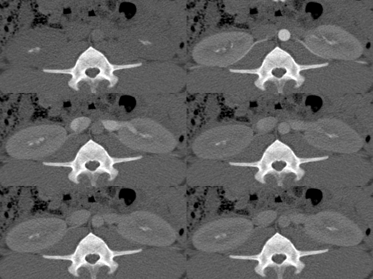
This gif shows a transit of a bolus of X-ray contrast media in the right and left kidneys of the animal model. These tomographic images were captured in a cross-sectional computed tomography (CT) scan of the abdomen and can be used to assess the function of the kidneys in kidney disease.

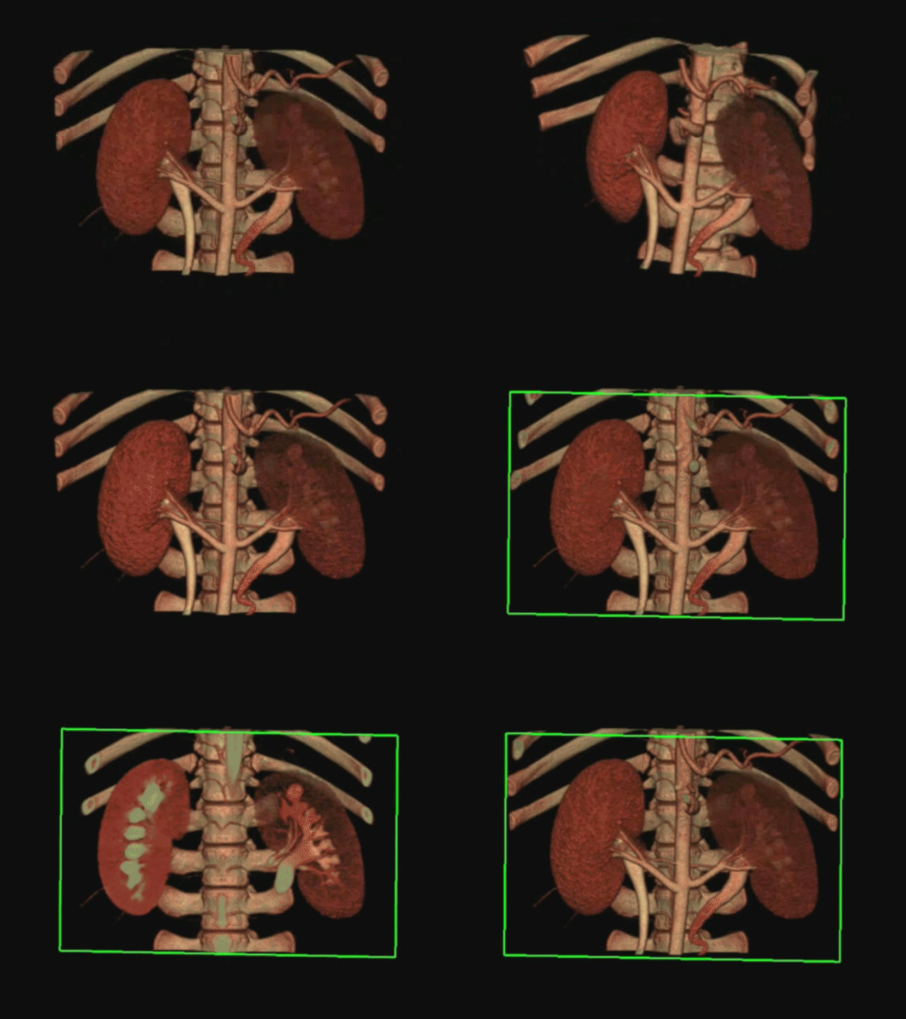
This gif shows in situ 3D reconstruction and tomographic sectioning of the kidneys from CT contrast-enhanced images. New image reconstruction techniques allow visualization of the kidneys in multiple angles and planes and thereby detection of subtle changes in kidney structure.

This gif shows the cardiac cycle visualized using CT contrast-enhanced images. Evaluation of the degree of contraction of the beating heart can be used to assess functional capacity in heart failure or regional changes after myocardial infarction.

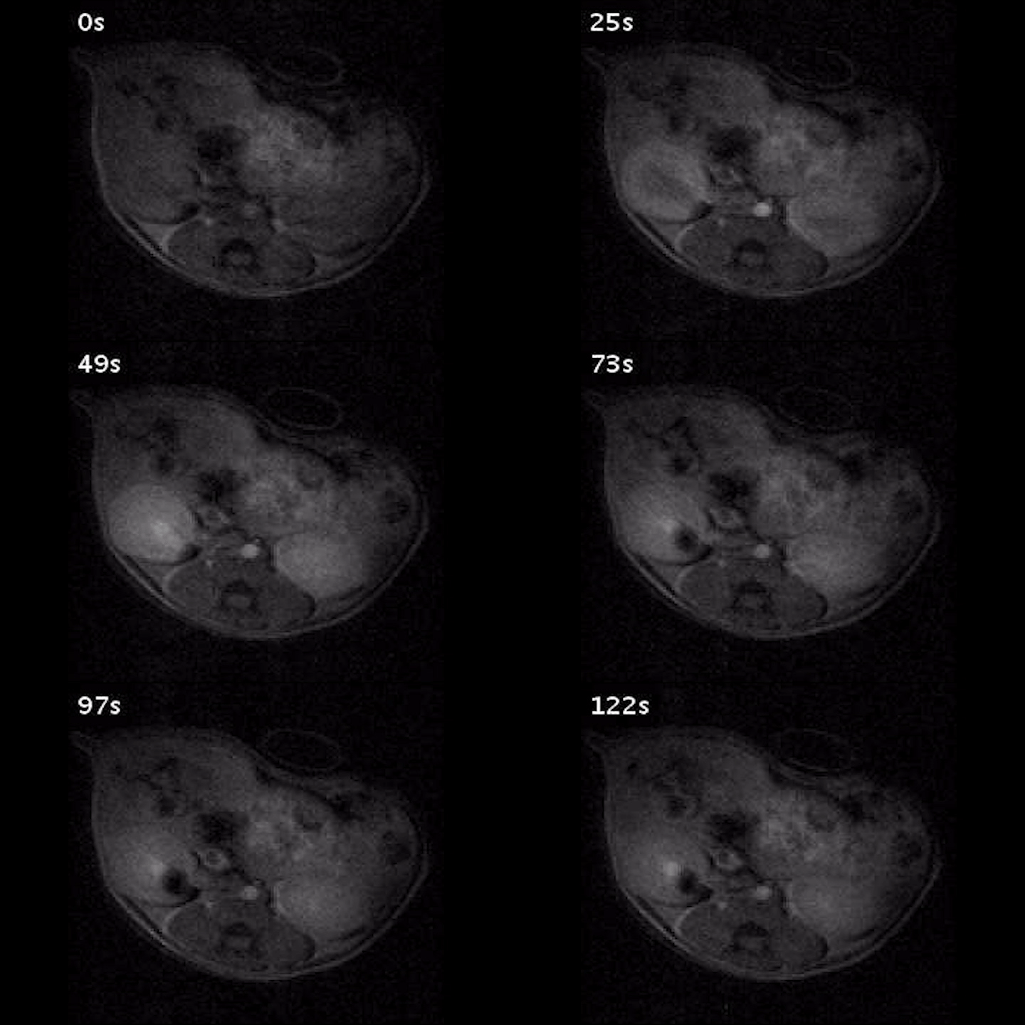
This gif shows the transit of a bolus of gadolinium in the right and left kidneys of an animal model detected by magnetic resonance imaging (MRI). Abdominal MRI provided noninvasive and relatively safe measurements of kidney function and structure in both small and large animal models, which are helpful in research of kidney disease.