-
Using sound waves to fight the progression of liver disease
Researchers in Dr. Ehman's Advanced Magnetic Imaging Technology Laboratory at Mayo Clinic use magnetic resonance elastography to determine the stiffness (fibrosis) of the liver without a biopsy.
Overview
The objective of this program is to expand the range of tissue, organ and system characteristics that can be noninvasively evaluated with magnetic resonance imaging (MRI) techniques. Research is directed at developing a novel "palpation by imaging" magnetic resonance technique to detect cancer and other tissue abnormalities at an early stage. The research uses MRI to directly image propagating acoustic waves in tissues, allowing images of viscoelastic properties to be generated.
The ongoing development of this technology has resulted in the first proven clinical application of magnetic resonance elastography (MRE): noninvasive detection of hepatic fibrosis. Chronic liver disease is a serious worldwide problem, and hepatic fibrosis is the most important consequence. Hepatic fibrosis can eventually lead to cirrhosis, which is irreversible and associated with high mortality. After extensive work to develop a method for reliably generating shear waves in the liver, the team developed a prototype technology for applying MRE to assess hepatic fibrosis. MRE-measured hepatic stiffness increases systematically with the fibrosis stage. Growing clinical experience indicates that MRE is at least as accurate as liver biopsy, while also being safer, more comfortable and less expensive.
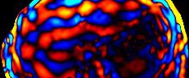
This research program is focused on developing technology and exploring the potential applications of MRE in assessing many other body systems. Studies have demonstrated that it is feasible to apply MRE to quantitatively assess the mechanical properties of skeletal muscle and tissue in the brain, thyroid, breast, myocardium, kidney, liver and skin. Among these applications, assessment of the brain has emerged as one of the most promising future clinical applications. The team's research has demonstrated promising results using these new biomarkers in preoperative assessment of meningiomas and skull base tumors and in assessment of neurodegenerative disease.
Affiliations
Dr. Ehman's Advanced Medical Imaging Technology Lab is affiliated with several Mayo Clinic research areas:
Resources
About Dr. Ehman
Dr. Ehman is a radiologist focused on body, musculoskeletal and cardiovascular magnetic resonance imaging (MRI). His research program develops methods to reduce or eliminate flow and tissue motion artifacts in MRI, new approaches for vascular imaging, and MRI-based techniques for characterizing the mechanical properties of tissue. He serves as a consultant in the Department of Physiology and Biomedical Engineering and as a professor of radiology in the Mayo Clinic College of Medicine and Science, Rochester, Minnesota.