Fluorescence in Situ Hybridization (FISH)
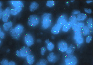
Locus-specific EGFR probe (orange) and centromere 7 probe (green): Not amplified
The Cytogenetics Core offers fluorescence in situ hybridization (FISH) services for a variety of specimen types, including nonparaffin, paraffin and paraffin tissue microarray.
FISH can be performed to enumerate the copy number of cancer-related genes and patterns of gene-related gains and losses; map the location of DNA sequences, genes or transgene insertions; and determine the presence or absence of interspecies cells in xenograft or chimeric animal models.
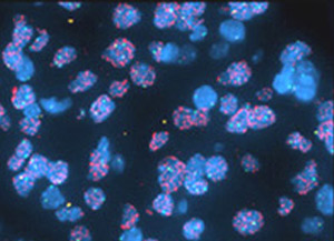
Locus-specific EGFR probe (orange) and centromere 7 probe (green): Amplified
Details
Nonparaffin FISH
Slides are processed and analyzed (counted) by the Cytogenetics Core, which includes image documentation.
- FISH is performed on interphase or metaphase cells that have been dropped on a slide or grown on a cover slip.
- Samples may be in the form of cultured cells or fresh or frozen tissue.
- Specific cell types include blood, bone marrow, tumors, cell lines, xenografts, hybrid cells from chimeric animal models, neural stem cells and mouse embryonic fibroblasts, among others.
Paraffin FISH
Slides are processed and analyzed by the Cytogenetics Core. Analysis includes image documentation.
- Paraffin-embedded tissue must be cut 5 micrometers thick and mounted on positively charged slides.
Paraffin FISH tissue microarray (TMA)
Paraffin-embedded tissue cores must be sectioned 5 micrometers thick and mounted onto a single slide.
- TMA slide processing service. Slides are processed (deparaffinized and set up) by the Cytogenetics Core.
- TMA analysis service. Slides are analyzed by the core on a "per sample core" basis. Analysis includes image documentation.
FISH imaging only
Slides are set up by the Cytogenetics Core or by the customer. Images are captured as the only result (no counting).
FISH probes
Inquire about commercially available probes:
- Centromere probes
- Gene-specific probes
- Locus-specific identifiers (LSI) probes
- Probe panels (several multicolor LSIs in one probe mix)
- Telomere probes
- Whole-chromosome paints
- Whole-genomic paints
The core also offers custom probe development.