Organs-on-Chip and Tissue Engineering
Researchers in the Cellular Microsystems and Biosensors Lab are pursuing multiple projects in the realm of tissue engineering and organ-on-chip. Examples include:
Microfluidic culture systems to model liver injury
Figure 1A
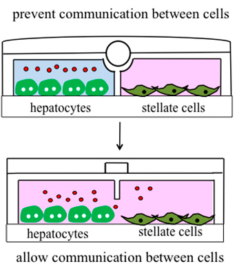
Figure 1A. Microfluidic co-cultures where communication between the cell types can be permitted or prevented through the use of valves.
The liver is composed of multiple liver cell types, including hepatocytes, hepatic stellate cells, macrophages, endothelial cells and cholangiocytes. Exposure to toxicants triggers complex fibrogenic cross-talk between the various cellular compartments, which leads to liver fibrosis and may result in liver failure. Addressing the questions of how levels of fibrogenic signals change over time and what cell types produce them may lead to the development of more-effective strategies for preventing or reversing liver fibrosis.
Approaches are being pursued by the laboratory to employ microfluidic culture systems (of the type shown in Figure 1A) where two or more cell types may be positioned within distinct but proximal microfluidic compartments. These microfluidic devices may be reconfigured by raising the wall or valve to either prevent or permit communication between neighboring cell compartments. When communication is prevented, a cellular compartment may be perturbed, for example, by exposure to an injurious agent. When communication is permitted, the injured cellular compartment is allowed to exchange paracrine signals with uninjured cells. Such systems may be used to study relative contributions of liver cell compartments to the milieu of fibrogenic signals associated with injury.
Designing microsystems for stem cell maintenance and differentiation
Figure 1B
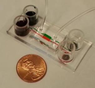
Figure 1B. An example of a microfluidic co-culture device used in the lab, with a penny shown for scale.
The laboratory has a long-standing interest in designing a microenvironment conducive to stem cell differentiation into hepatocytes. For example, researchers have employed growth factor immobilization on culture surfaces to signal to stem cells from the bottom up and drive hepatic differentiation of stem cells. More recently the lab is using microfluidic cultures for optimizing stem cell differentiation protocols and harnessing endogenous signals from stem cells.
Devices used for stem cell cultivation are similar to those shown in Figure 1B. When cultured in such devices, mouse embryonic stem cells maintain pluripotency in the absence of soluble leukemia inhibitory factor (LIF) — a pluripotency inducing signal that is typically present in stem cell media in copious amounts. This effect is due to the fact that stem cells in microfluidic chambers produce endogenous LIF.
Efforts are currently under way to demonstrate similar accumulation of endogenous signals in human pluripotent stem cells. In addition, the lab is developing stem cell differentiation strategies by creating microfluidic co-culture systems where adult hepatocytes are maintained in chambers adjacent to stem cells in order to drive hepatic differentiation of stem cells.
Key publications
Zhou Q, Patel D, Kwa T, Haque A, Matharu Z, Stybayeva G, Gao Y, Diehl AM, Revzin A. Liver injury-on-a-chip: Microfluidic co-cultures with integrated biosensors for monitoring liver cell signaling during injury. Lab on a Chip. 2015;23:4467.
Haque A, Gheibi P, Gao Y, Foster E, Son KJ, You J, Stybayeva G, Patel D, Revzin A. Cell biology is different in small volumes: Endogenous signals shape phenotype of primary hepatocytes cultured in microfluidic channels. Scientific Reports. 2016;6:33980.
Guild J, Haque A, Gheibi P, Gao Y, Son KJ, Foster E, Dumont S, Revzin A. Embryonic stem cells cultured in microfluidic chambers take control of their fate by producing endogenous signals including LIF. Stem Cells. 2016;34:1501.
Haque A, Gheibi P, Stybayeva G, Gao Y, Torok N, Revzin A. Ductular reaction-on-a-chip: Microfluidic co-cultures to study stem cell fate selection during liver injury. Scientific Reports. 2016;6:36077.
Microencapsulation of cells
Figure 2A

Figure 2A. Microfluidic device for fabricating microcapsules.
Figure 2B
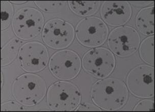
Figure 2B. Example of 400-mm diameter microcapsules carrying hepatocyte spheroids. A similar strategy is being used to encapsulate islets and stem cells.
Translation and scaling-up of stem cell therapies may benefit from the development of microcapsules that will serve as cell carriers during expansion and differentiation in vitro, and as vehicles for stem cell transplantation. The research team has been employing microfluidics to fabricate cell-carrying hydrogel microcapsules of uniform diameter and cell number. We have demonstrated that both stem cells and adult cells may be encapsulated and maintained within these hydrogel carriers without loss of viability or function. Experiments are currently underway to encapsulate human pluripotent stem cells and differentiate these cells toward hepatocytes or pancreatic beta cells.
Key publications
Siltanen C, Diakatou M, Lowen J, Haque A, Rahimian A, Stybayeva G, Revzin A. One step fabrication of hydrogel microcapsules with hollow core for assembly and cultivation of hepatocyte spheroids. Acta Biomaterialia. 2017;50:428.
Siltanen C, Yaghoobi M, Haque A, You J, Lowen J, Soleimani M, Revzin A. Microfluidic fabrication of bioactive microgels for rapid formation and enhanced differentiation of stem cell spheroids. Acta Biomaterialia. 2016;34:125.